Brain MRI: What to Expect Before, During, and After the Scan

A brain MRI is a noninvasive imaging test that creates detailed pictures of the brain and nearby structures. Knowing what happens before, during, and after the scan can help patients feel more prepared and comfortable.
Overview of a Brain MRI
A brain MRI, or magnetic resonance imaging scan, is a test that produces highly detailed pictures of the brain, skull base, blood vessels, nerves, and nearby soft tissues. It does not use X-rays. Instead, it uses a strong magnetic field and radio waves to generate images that can help doctors understand symptoms and guide treatment decisions.
Doctors may request a brain MRI to investigate headaches, dizziness, seizures, weakness, vision changes, hearing problems, memory concerns, or changes in balance. It may also be used to monitor known conditions, plan surgery, or check recovery after treatment. In many cases, it provides more detail than other imaging tests for soft tissues in the brain.
A brain MRI can help assess a wide range of concerns, including stroke, multiple sclerosis, infections, inflammation, tumors, bleeding, structural changes, or signs of neurodegenerative disease. It may also be part of the evaluation for conditions such as brain tumors or stroke. The scan itself is usually painless, and most people go home shortly after it is completed.
Why a Brain MRI May Be Recommended
A clinician may recommend a brain MRI when symptoms suggest a problem affecting the brain or nervous system. Compared with many other imaging methods, MRI is especially useful for showing fine details of the brain tissue, fluid spaces, and surrounding structures. This makes it valuable when symptoms are not fully explained by an exam alone.
Common reasons for a brain MRI include ongoing or severe headaches, fainting, seizures, confusion, numbness, weakness, personality changes, or unexplained vision and hearing symptoms. It may also be used after a head injury, to evaluate possible infection, or to monitor known findings over time. In children and adults alike, MRI can help doctors look for causes without exposing the patient to radiation.
Sometimes a brain MRI is performed with a special dye called contrast, usually gadolinium-based contrast, which is injected into a vein. Contrast can help highlight inflammation, certain tumors, blood vessels, and areas where the blood-brain barrier is disrupted. In selected cases, the scan may be combined with advanced techniques or related imaging such as MRI imaging or neuroradiology evaluation.
How to Prepare Before the Scan

Preparation for a brain MRI is often simple, but it is important to follow the imaging center’s instructions. Patients are usually asked to remove jewelry, hairpins, hearing aids, watches, removable dental work, and other metal items before entering the MRI room. Because the magnet is very strong, even small metal objects can be unsafe or interfere with image quality.
Before the scan, the care team will ask about medical history, especially any pacemaker, aneurysm clips, cochlear implants, metal fragments, implanted pumps, artificial joints, or prior surgeries. Many modern implants are MRI-compatible, but each device must be checked carefully. Patients should also mention pregnancy, kidney disease, dialysis, or a past reaction to contrast material.
If contrast is planned, some patients may need a blood test to assess kidney function, depending on their health history. Eating and drinking are often allowed, although some centers may give different instructions if sedation is planned. People who feel anxious in enclosed spaces should tell their doctor in advance, as relaxation techniques or mild medication may sometimes be considered.
Comfortable clothing without metal fasteners may be allowed, but many patients change into a hospital gown. It can also help to arrive a little early to complete safety forms and ask questions. Understanding the steps beforehand often makes the experience feel easier and more predictable.
What Happens During a Brain MRI
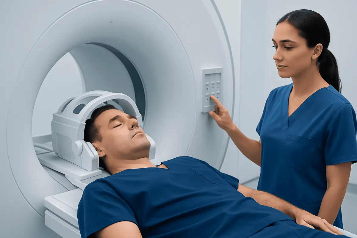
During a brain MRI, the patient lies on a narrow table that slides into the scanner. The head may rest in a special support or coil that helps produce clear images. The technologist positions the patient carefully and explains how to stay still, since movement can blur the pictures and may require parts of the scan to be repeated.
Once the scan begins, the machine makes loud tapping, knocking, or humming sounds as it collects images. Earplugs or headphones are usually provided for comfort. The technologist watches from a nearby control room and can communicate through an intercom, so the patient is not left without support.
The most important part of the scan is remaining as still as possible. A brain MRI may take about 20 to 60 minutes, depending on the reason for the test and whether contrast or additional sequences are needed. Some people find the enclosed space uncomfortable, but many complete the scan well with reassurance, breathing exercises, and clear guidance from staff.
If contrast is required, it is usually injected through a small IV during part of the exam. The injection may feel cool in the arm, but it is generally well tolerated. In some situations, the MRI may be part of a broader work-up that includes a comprehensive brain check-up to assess neurological symptoms in a structured way.
What to Expect After the Scan and When Results Arrive
After a brain MRI, most patients can return to normal activities right away. If no sedation was used, there is usually no recovery period beyond getting dressed and receiving any practical instructions. If contrast was given, drinking fluids afterward may be encouraged unless a doctor has advised fluid restriction for another medical reason.
When sedation or anti-anxiety medicine has been used, the patient may need monitoring for a short time and should usually avoid driving until fully alert. A family member or friend may be asked to accompany them home. The imaging team will explain any temporary precautions that apply.
The images are reviewed by a radiologist, a doctor trained to interpret scans. The radiologist sends a report to the referring clinician, who then discusses the findings with the patient in the context of symptoms, examination, and any other tests. Some results are available quickly, but timing varies depending on the urgency and the healthcare setting.
It is important to remember that a brain MRI report may describe both significant findings and minor incidental changes. Not every abnormality is dangerous, and many findings need to be interpreted alongside age, medical history, and current symptoms. Patients who have questions should ask their doctor to explain the report in clear, practical terms.
Safety, Risks, and Special Situations
Brain MRI is considered very safe for most people because it does not use ionizing radiation. However, MRI safety depends on careful screening for metal devices or fragments that could move, heat up, or stop working properly in the magnetic field. This is why the pre-scan safety checklist is such an important part of the process.
Contrast-enhanced MRI is also safe for many patients, but it is not suitable for everyone. People with severe kidney problems may need extra assessment before receiving gadolinium-based contrast. Although allergic-type reactions are uncommon, patients should tell the team about any previous contrast reaction or other significant allergies.
Claustrophobia can also be a challenge for some patients. Helpful strategies include asking for a clear explanation before the scan, practicing slow breathing, listening to music if available, or discussing sedation options in advance. Children or adults who have trouble staying still may sometimes need additional support to complete the exam successfully.
Pregnant patients should inform their doctor and the MRI team. In many cases, MRI can still be appropriate during pregnancy when medically needed, but the decision should be individualized. A healthcare professional will weigh the benefits of the information gained against any potential concerns based on the person’s situation.
When to Contact a Doctor and Practical Tips
Most people have no problems after a brain MRI, but patients should seek medical advice if they develop symptoms such as rash, swelling, breathing difficulty, severe dizziness, or persistent vomiting after contrast injection. These symptoms are uncommon, but they deserve prompt assessment. Patients should also contact their doctor if they have pain, redness, or swelling at the IV site that worsens over time.
It is also reasonable to follow up if results are delayed or if symptoms change while waiting for the report. New weakness, trouble speaking, seizures, sudden severe headache, confusion, or loss of consciousness need urgent medical attention, whether or not a scan has recently been done. The MRI is one part of care, and new symptoms should never be ignored.
Practical tips can make the day easier:
- Bring a list of implants, surgeries, and current medications.
- Arrive early to complete screening forms calmly.
- Wear simple clothing and avoid metal accessories.
- Tell the staff right away about anxiety, pain, pregnancy, or difficulty lying flat.
- Ask when and how results will be shared before leaving the imaging center.
For patients seeking coordinated neurological imaging and follow-up, Acıbadem Health Point’s multidisciplinary specialists and JCI-accredited hospitals diagnose and treat brain conditions for international patients. Clear communication before and after imaging can help patients feel informed and supported throughout the process.
Frequently asked questions
01Is a brain MRI painful?
A brain MRI is usually painless. The patient lies still on a table while the machine takes images, but there are no needles unless contrast is needed. Some people find the noise or enclosed space uncomfortable rather than painful.
02How long does a brain MRI take?
Many brain MRI scans take about 20 to 60 minutes. The exact length depends on whether contrast is used and how many image sequences are needed. More specialized studies may take longer.
03Can a patient eat before a brain MRI?
Often yes, but instructions can vary. If sedation or contrast is planned, the imaging center may give specific advice about food and drink. It is best to follow the instructions provided for that appointment.
04What should not be worn to a brain MRI?
Metal items should not be worn into the MRI room. This includes jewelry, watches, hair accessories with metal, hearing aids, and clothing with certain metal fasteners. The imaging staff will explain what must be removed.
05Why would contrast be used in a brain MRI?
Contrast can help highlight certain tissues and blood vessels more clearly. It is often used when doctors need more detail about tumors, inflammation, infection, or areas of abnormal blood flow. Not every brain MRI requires contrast.
06Is a brain MRI safe for people with implants?
Many people with implants can still have an MRI, but the specific device must be checked first. Some implants are MRI-compatible under certain conditions, while others may prevent scanning. Patients should always tell the MRI team about any implant or prior surgery.
This article is for general information only and is not a substitute for professional medical advice. Please consult a qualified doctor about your individual situation.
Explore treatments in Turkey — costs, top hospitals & a free quote
JCI-accredited · board-certified surgeons · reply within 24h
Add us as a Preferred Source to see more of our trusted health content across Google Search, AI Overviews and Discover.