Brain CT Scan for Bleeding, Stroke, and Head Injury
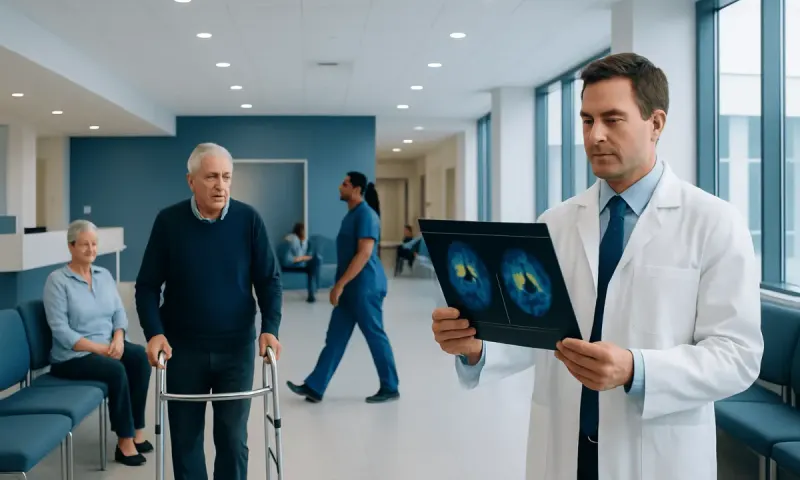
A CT scan brain, also called a brain or head CT, is a quick imaging test that creates detailed cross-sectional pictures of the brain and skull. It is commonly used after head injury or sudden neurological symptoms because it can rapidly identify bleeding, major swelling, fractures and some types of stroke.
CT Scan Brain: What It Is and Why It Is Used
A CT scan brain uses a series of X-rays and computer processing to create detailed images of the brain, skull, sinuses and surrounding tissues. CT stands for computed tomography. Unlike a standard X-ray, it produces slice-like images that allow clinicians to assess structures inside the head more closely.
The test is often requested when a clinician needs information quickly. It can help assess a recent head injury, sudden severe headache, confusion, weakness, speech difficulty, seizure, loss of consciousness or suspected stroke. It may also be used to monitor certain findings over time or to assess the brain after an operation.
A brain CT does not replace a full neurological assessment. The scan is interpreted alongside symptoms, examination findings, medical history and, when needed, other tests such as blood tests, MRI or vascular imaging. The most appropriate imaging study depends on the person’s symptoms and how urgently answers are needed.
How the Brain CT Procedure Works
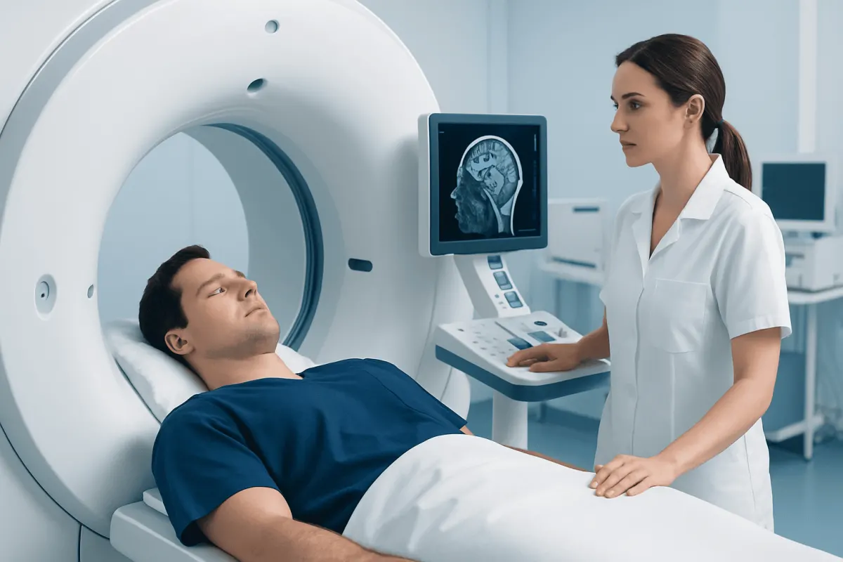
Before the scan, the person is usually asked to remove metal objects around the head and neck, such as glasses, hearing aids, hair clips, removable dental appliances or jewellery. They lie on a padded table, generally on their back, with the head positioned to help keep it still. The table then moves through the short, ring-shaped CT scanner.
The scanner does not normally touch the person, and the radiographer can communicate throughout the examination. Remaining still is important because movement can blur the images. The machine may make quiet whirring sounds, but a CT scan is usually less enclosed than an MRI scan.
Some examinations use intravenous contrast, a dye that makes blood vessels, inflammation, tumors and certain tissues easier to see. A brief warm sensation or metallic taste can occur when contrast is injected and usually passes quickly. The imaging team should be told about previous contrast reactions, kidney disease, pregnancy, diabetes, thyroid conditions and all regular medicines.
Administrative labels such as a CT scan brain procedure code may appear on referrals, billing documents or medical records. The exact code varies by healthcare system and whether contrast is used, so the imaging provider or insurer is the appropriate source for coding questions.
How Long Does a CT Scan of the Brain Typically Take?
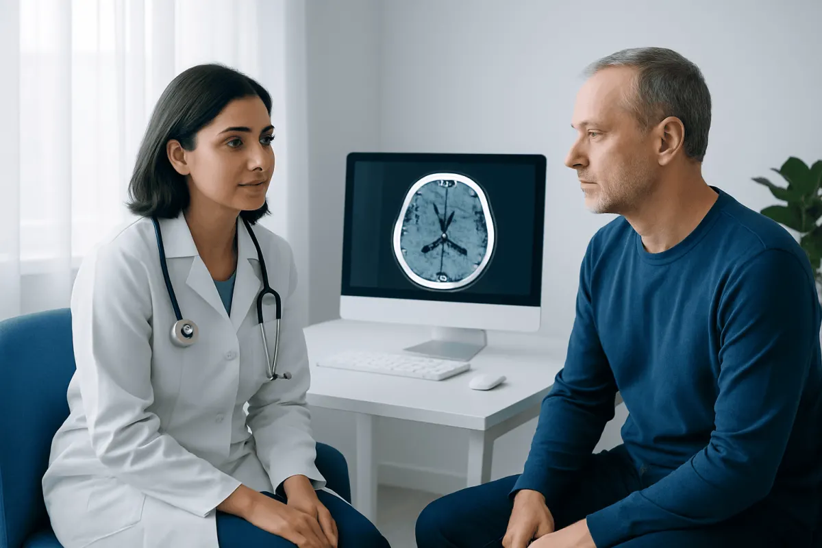
The actual image acquisition for a brain CT scan often takes less than a minute. From entering the scan room to completion, many routine examinations take about 10 to 20 minutes. More time may be needed for registration, safety checks, positioning, contrast administration or additional images.
In an emergency department, the scan itself can often be arranged promptly when clinicians are concerned about conditions such as head bleeding or stroke. However, waiting time before the scan may vary according to the urgency of the situation, the number of patients needing imaging and local service arrangements.
People who are in pain, have difficulty lying flat, are very anxious or cannot remain still may need extra support. Children and some adults may occasionally require sedation, but this is not routine and requires specific preparation and aftercare instructions.
What Are the Common Abnormalities Found on a Brain CT Scan?
A head CT is particularly sensitive for fresh bleeding in and around the brain. It can identify bleeding after trauma, bleeding caused by a ruptured blood vessel, and some bleeding related to stroke. It can also show skull fractures, collections of blood outside the brain, brain swelling and pressure effects that may require urgent assessment.
Other common findings can include changes from an established stroke, enlarged fluid spaces in the brain, hydrocephalus, calcifications, sinus disease and age-related changes. CT may show a mass or an area that needs further evaluation, although MRI often provides greater detail for many tumors, inflammation, subtle injuries and abnormalities of the brain tissue.
A report may also describe findings that are unrelated to the reason for the scan. Many incidental findings are harmless or need only routine follow-up, but their importance depends on the individual context. A treating clinician should explain what a result means and whether further assessment is needed.
After a significant head injury, CT findings may help guide care for traumatic brain injury, but a normal scan does not always exclude concussion. Concussion is primarily diagnosed from symptoms and clinical assessment, and some symptoms may develop or change after the initial scan.
Can a CT Scan Detect Anything in the Brain?
Yes. A CT scan can detect many important changes in the brain and skull, especially those that need rapid assessment. These include acute bleeding, major stroke-related changes, skull fractures, swelling, hydrocephalus, large masses and some infections or inflammatory changes.
However, a normal CT scan does not rule out every neurological condition. Very early ischemic stroke, small lesions, subtle traumatic changes, some infections, multiple sclerosis and many causes of headache or dizziness may not be clearly visible. In these situations, a clinician may recommend MRI, repeat imaging, blood tests or Stroke Specialist Evaluation: What to Expect at Your First Vascular Neurology Visit" class="ahp-ilk">specialist evaluation.
For suspected stroke, CT is commonly the first imaging test because it is fast and helps distinguish bleeding from other causes of symptoms. Additional scans may be used to examine brain blood vessels or blood flow. Anyone with sudden facial drooping, arm weakness, speech difficulty, severe balance problems or sudden vision loss should seek emergency help immediately.
Preparing for a Head CT and What Not to Do Before It
For a non-contrast head CT, there is usually little special preparation. People should follow the instructions given by the imaging center and bring relevant previous imaging reports or scan discs if requested. It is helpful to arrive on time and tell the team about implants, previous surgery, allergies and current symptoms.
What should you not do before a head CT scan? Do not wear metal objects that cannot be removed, and do not withhold prescribed medicines unless a clinician specifically advises this. For a scan involving contrast, the imaging service may ask the person not to eat for a short period beforehand; instructions differ, so patients should not fast unless they have been told to do so.
It is important not to overlook information about possible pregnancy, past contrast reactions or kidney problems. These details do not automatically prevent CT imaging, particularly when an urgent scan is needed, but they help the care team choose the safest approach. People with diabetes or those taking medicines that affect kidney function should ask whether any individual preparation is required.
Recovery and How Long It Takes to Get Brain CT Results
Most people have no recovery period after a CT scan and can eat, drink, drive and return to normal activities straight away. Drinking fluids after contrast may be advised unless a clinician has recommended fluid restriction for another health condition. A delayed allergic-type reaction to contrast is uncommon, but new rash, itching, breathing difficulty or swelling should be assessed urgently.
How long to get brain CT scan results depends on why and where the scan is performed. In an emergency setting, the images may be reviewed promptly by a radiologist and treating team, sometimes within minutes to hours. For outpatient examinations, a formal report may be sent to the referring clinician within a few days, depending on local processes.
CT scan results after concussion are often normal because concussion can affect brain function without causing visible bleeding or structural injury. Clinicians use the result together with symptoms such as headache, dizziness, nausea, concentration problems or sleep changes. Worsening symptoms after head injury need urgent reassessment even if an earlier scan was reassuring.
A CT scan after brain surgery may be ordered soon after the procedure or during follow-up to check for bleeding, swelling, fluid collections, air, drain position or other postoperative changes. The surgical team will explain the expected findings and what follow-up imaging, if any, is appropriate.
When to Seek Medical Care
Emergency medical care is needed after a head injury when there is loss of consciousness, repeated vomiting, increasing drowsiness, seizure, worsening headache, confusion, weakness, numbness, unequal pupils, trouble speaking, difficulty walking or fluid or blood leaking from the nose or ears. This is especially important for older adults, people taking blood-thinning medicines and anyone with a bleeding disorder.
Urgent assessment is also appropriate for sudden neurological symptoms, including a severe new headache unlike previous headaches, facial drooping, weakness on one side, speech or vision changes, fainting, new seizure or sudden confusion. These symptoms can have several causes, and prompt evaluation helps clinicians determine whether brain imaging is needed.
For non-urgent concerns, a doctor can review persistent headaches, changes in memory, ongoing dizziness, new balance issues or symptoms that continue after concussion. Acıbadem Health Point’s multidisciplinary specialists and JCI-accredited hospitals can assess neurological symptoms and arrange appropriate imaging and follow-up for international patients.
Frequently asked questions
01Is a CT scan of the brain painful?
A brain CT scan is not painful. The person needs to lie still on a table while it moves through the scanner. If contrast is used, there may be a brief warm feeling or metallic taste during the injection.
02Do I need contrast for a CT scan brain?
Not always. A non-contrast scan is often used first for acute head injury, suspected bleeding and many stroke assessments. Contrast may be added when clinicians need a clearer view of blood vessels, inflammation, infection or a suspected mass.
03Can a normal brain CT rule out a concussion?
No. A CT scan is useful for identifying bleeding, fractures and other structural injuries, but concussion may not appear on CT images. Diagnosis is based on the injury history, symptoms and neurological examination.
04Is brain CT radiation dangerous?
CT uses ionizing radiation, and imaging teams aim to use the lowest dose needed to answer the clinical question. When a CT is medically indicated, its ability to identify urgent problems generally outweighs the small radiation risk. Patients can ask their clinician why the scan is recommended and whether another test would be suitable.
05Can I eat and drink before a head CT?
For many non-contrast head CT scans, people can eat and drink normally. If contrast or sedation is planned, the imaging center may provide fasting instructions. It is best to follow the specific directions given for the appointment.
06What happens if the brain CT shows an abnormality?
The treating clinician will explain the finding in the context of the person’s symptoms and medical history. Some findings require urgent treatment, while others need further imaging, follow-up or no treatment at all. A radiology report alone should not be used to self-diagnose.
This article is for general information only and is not a substitute for professional medical advice. Please consult a qualified doctor about your individual situation.
Hair transplant in Turkey — costs, graft counts & a free assessment
JCI-accredited · board-certified surgeons · reply within 24h
Add us as a Preferred Source to see more of our trusted health content across Google Search, AI Overviews and Discover.