


Testicular Dermoid Cyst Causes and Treatment
Testicular Dermoid Cyst Causes and Treatment Testicular dermoid cysts are uncommon benign growths that can develop within the testes. These cysts are a type of germ cell tumor composed of mature tissues such as skin, hair follicles, and sometimes even sebaceous glands, all encapsulated within a cystic structure. While dermoid cysts are more frequently associated with the ovaries, their presence in the testes is rare but noteworthy due to their potential implications and the need for accurate diagnosis and management.
The exact cause of testicular dermoid cysts remains unclear. They are believed to originate from remnants of embryonic germ cells that fail to differentiate properly during fetal development. These pluripotent cells can give rise to various tissue types, which explains the diverse components found within dermoid cysts. Genetic factors may play a role, but no specific hereditary patterns have been conclusively identified. Additionally, some theories suggest that developmental anomalies during embryogenesis lead to the formation of these cysts. Unlike malignant tumors, dermoid cysts are benign, but their growth can sometimes cause discomfort or swelling, prompting medical evaluation.
In terms of causes, it is crucial to distinguish between congenital origins and acquired factors. Congenital development is the primary suspected cause, as these cysts are often discovered in young males during routine examinations or investigations for painless testicular swelling. Trauma or infections are not typically associated with dermoid cyst formation, although they may complicate the clinical picture if present. It is important to note that while dermoid cysts are benign, they can sometimes be mistaken for malignant testicular tumors based on imaging studies, which underscores the importance of thorough diagnostic procedures.
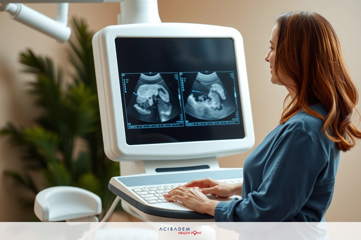
Diagnosis often involves a combination of physical examination, imaging studies, and histopathological analysis. During a physical exam, a firm, painless mass may be palpable within the testicle. Ultrasound imaging is the primary modality used to evaluate testicular masses, revealing characteristic features such as well-defined cystic areas with mixed echogenicity. Sometimes, magnetic resonance imaging (MRI) may be employed to better delineate the cyst’s content and extent. However, definitive diagnosis usually requires surgical removal of the cyst followed by histopathological examination, which confirms the presence of mature tissue components characteristic of dermoid cysts.
Treatment for testicular dermoid cysts primarily involves surgical excision. The goal is to remove the cyst completely while preserving as much healthy testicular tissue as possible, especially in young patients concerned with fertility. A common surgical approach is testis-sparing surgery, which involves enucleation of the cyst through an inguinal incision. This procedure minimizes the risk of damaging surrounding structures and allows for tissue preservation. In some cases, if the cyst is extensive or suspicion of malignancy persists, orchiectomy—the removal of the entire testicle—may be necessary. Postoperative follow-up includes regular imaging to monitor for recurrence, though it is rare with complete excision. Prognosis after removal is excellent, as dermoid cysts are benign and do not tend to recur if fully excised.
In summary, testicular dermoid cysts are rare benign growths stemming from embryonic tissue remnants. Although their exact causes are not entirely understood, they are generally congenital in origin. Accurate diagnosis relies on imaging and histopathology, and treatment involves surgical removal with an emphasis on preserving testicular function. Early detection and management ensure excellent outcomes and help alleviate symptoms or concerns related to testicular masses.


