


Understanding ASL for Carotid Cavernous Fistulas
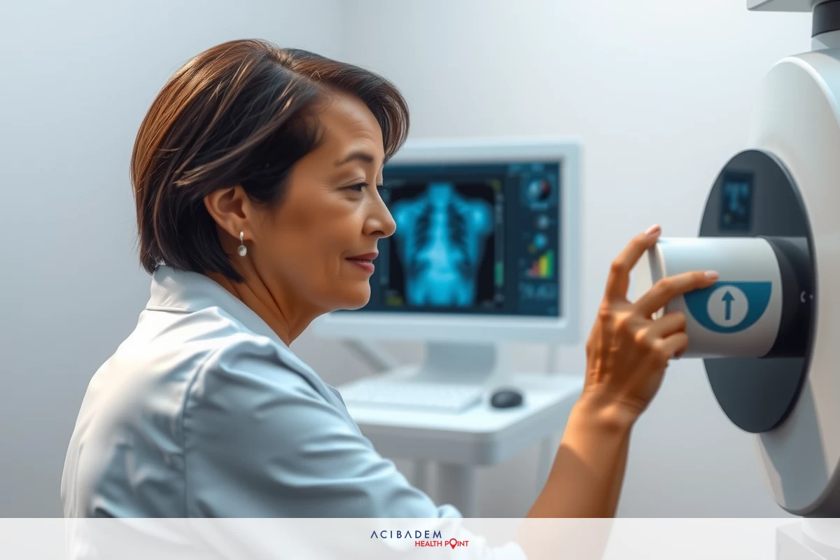
Understanding ASL for Carotid Cavernous Fistulas Arterial Spin Labeling (ASL) is an advanced brain imaging technique that enables non-invasive diagnosis of Carotid Cavernous Sinus Fistula (CCF). Utilizing a specialized MRI approach, it detects abnormal blood flow in the brain without the need for surgery.
Early and accurate detection of CCF is crucial. ASL MRI provides clear blood flow imaging without requiring contrast dye, making the procedure safer for patients. Understanding ASL for Carotid Cavernous Fistulas
Let’s explore how ASL aids in detecting and evaluating Carotid Cavernous Sinus Fistulas and why it’s an valuable imaging tool today.
What is a Carotid-Cavernous Fistula?
Carotid cavernous fistula (CCF) is a serious condition involving the eyes and brain. Understanding its nature, causes, and symptoms is crucial.
Certainly! Please provide the original text you’d like me to rewrite.
Understanding ASL for Carotid Cavernous Fistulas A carotid-cavernous fistula (CCF) is an abnormal link between the carotid arteries and the cavernous sinus—a large vein at the brain‘s base. This abnormal connection disrupts normal blood flow, increases intracranial pressure, and can damage surrounding brain structures.
Causes
CCF may occur due to several reasons.
- Trauma: Head injuries from blows or cuts can frequently lead to CCF.
- Spontaneous Onset: Certain conditions, such as connective tissue disorders, can cause CCF without trauma.
- Surgical complications involving the skull or blood vessels may occasionally cause CCF.
Signs and Symptoms
The symptoms of CCF depend on the severity and duration of the condition, with common indicators including:
- Elevated intraocular pressure can cause eye pain and potentially result in vision loss.
- Diplopia: Double vision caused by misaligned eyes due to nerve damage.
- Proptosis: An abnormal protrusion of the eye, often pulsating, indicating exophthalmos.
| Cause | Description |
|---|---|
| Trauma | Head injuries leading to CCF. |
| Spontaneous Development | Conditions like connective tissue disorders causing CCF without trauma. |
| Surgical Complications | CCF resulting from surgeries involving the skull base or vascular system. |
Grasping the Basics of ASL Imaging
Arterial Spin Labeling (ASL) is an MRI technique that measures brain blood flow without using contrast agents, making it a safe option for applications such as stroke assessment.
Core Principles
ASL uses magnets to label blood water, then compares the labeled and unlabeled images to assess blood flow, allowing us to evaluate brain circulation.
Understanding How ASL Functions
ASL employs MRI to tag blood in arteries prior to reaching the brain. This labeled blood disperses similarly to a contrast agent. By capturing images before and after labeling, we can assess blood flow efficiency. Understanding ASL for Carotid Cavernous Fistulas
Advantages of Using ASL in Medical Imaging
ASL doesn’t require contrast agents, making it safe for individuals with allergies or kidney issues. Its repeatability allows doctors to monitor blood flow and treatment progress over time, providing a reliable and safe imaging method.
“ASL Imaging for Carotid-Cavernous Fistulas”
Arterial Spin Labeling (ASL) is an innovative MRI technique for diagnosing Carotid Cavernous Fistulas (CCF). It safely visualizes and quantifies cerebral blood flow by using the body’s own blood as a natural tracer.
ASL effectively detects blood flow issues in CCF, highlighting the increased flow typical of these fistulas. It’s advantageous because it doesn’t require contrast dye, making it suitable for patients with dye allergies or sensitivities.
ASL assists doctors in better understanding blood flow by analyzing both incoming and outgoing blood. This comprehensive view helps in accurately diagnosing issues and determining the most effective treatment plan.
Below is a table comparing ASL to other MRI methods for CCF:
| Feature | ASL | Traditional MRI |
|---|---|---|
| Contrast Requirement | None ( Endogenous tracer ) | Required (Exogenous contrast agents) |
| Invasiveness | Non-invasive | Moderately invasive (with contrast) |
| Flow Quantification | Direct ( Quantitative perfusion ) | Indirect (Dependent on contrast dynamics) |
| Kidney Safety | Safe (No nephrotoxic agents) | Risk in patients with renal impairment |
Utilizing ASL in brain imaging, particularly for CCF, marks significant progress by providing safer, more precise blood flow assessment. This improves doctors’ ability to
develop effective treatment strategies.
Benefits of Using ASL in Fistula Detection
Arterial Spin Labeling (ASL) offers significant diagnostic benefits for detecting carotid cavernous fistulas. As a non-invasive technique, it produces detailed images without requiring contrast agents, unlike traditional digital subtraction angiography that relies on injected contrast. Understanding ASL for Carotid Cavernous Fistulas
ASL reduces radiation exposure by using MRI instead of ionizing radiation, ensuring patients, especially children and those sensitive to radiation, avoid unnecessary radiation during multiple tests.
ASL provides detailed images that enable doctors to diagnose accurately. It visualizes blood flow, aiding in the detection and assessment of fistulas. This supports effective treatment planning and monitoring.
ASL is simple to implement in hospitals since it integrates seamlessly with existing equipment, avoiding costly upgrades or major adjustments. This makes it an economical and practical option for healthcare providers.
| Advantages | ASL | Traditional Imaging |
|---|---|---|
| Non-invasive Method | ✔ | ✖ |
| Radiation Exposure Reduction | ✔ | ✖ |
| Follow-up Evaluations | ✔ | Limited due to radiation risk |
| High-resolution Imaging | ✔ | ✔ |
| Cost-effective | ✔ | Variable |
ASL is an effective and safe method for detecting carotid cavernous fistulas, with minimal radiation exposure. It’s useful for ongoing patient monitoring and enhances neuroimaging capabilities when integrated into hospital settings.
“Neuroimaging Applications of ASL in Clinical Practice”
Arterial Spin Labeling (ASL) is an innovative imaging technique that allows clinicians to assess cerebral blood flow, including in conditions like Carotid Cavernous Fistulas (CCFs).
‘Accuracy of Diagnosis’
ASL effectively detects Carotid Cavernous Fistulas by providing a non-invasive, direct measurement of blood flow, ensuring high accuracy.
ASL distinctly visualizes blood vessels without the need for contrast dye, unlike conventional imaging techniques.
‘Comparison with Alternative Imaging Methods’
ASL differs from CT, MRI, and traditional angiography. Unlike traditional angiography, which provides detailed images but involves invasiveness and radiation, ASL is a safer, non-radiative alternative.
Using ASL with MRI can be advantageous in certain situations, as it leverages the strengths of both techniques to improve diagnosis.
Here’s a chart comparing ASL to other imaging techniques.
| Imaging Technique | Invasiveness | Radiation Exposure | Contrast Agents | Sensitivity | Specificity |
|---|---|---|---|---|---|
| ASL | Non-Invasive | None | Not Required | High | High |
| Traditional Angiography | Invasive | High | Required | Very High | Very High |
The table indicates that ASL is an effective and safe option for brain imaging. The ideal imaging technique varies by case, but ASL offers a balanced combination of accuracy and patient safety. Understanding ASL for Carotid Cavernous Fistulas
Recent Studies on ASL Imaging for Carotid-Cavernous Fistulas
Recent advances in medical research have enhanced the use of Arterial Spin Labeling (ASL) for diagnosing carotid cavernous fistulas. This review highlights current studies and explores future prospects for ASL in this field.
Recent Research
Recent research highlights ASL’s ability to produce detailed images of carotid cavernous fistulas, emphasizing its accuracy and reliability in detecting these complex vascular issues. For instance, a study in the *American Journal of Neuroradiology* demonstrated that ASL provides clear images without requiring contrast dye.
Researchers at Stanford University compared ASL with traditional imaging techniques and discovered that ASL more effectively detects issues and enhances patient comfort. This highlights the crucial role of research in improving imaging methods.


